Digestive oncology
ESD - Endoscopic Submucosal Dissection (Esophagus/Stomach)
This procedure uses an endoscope to safely remove superficial lesions in the lining of the esophagus or stomach without surgery or scarring.
What is it?
This is a procedure that uses an endoscope (a flexible tube that can be inserted into the esophagus) to remove lesions confined to the mucous membrane. In the past, such lesions were removed surgically. With this new technique, the lesions can be safely removed without scarring and without surgery, resulting in a shorter hospital stay.
These are usually lesions that (if left untreated) could become malignant or are already malignant but have not yet invaded beyond the mucosa. In most cases, an endoscopic ultrasound (EUS) examination will be performed beforehand to confirm the depth. This technique is used for the removal of larger lesions so that they can be removed in one piece.
Please report the following during the preliminary consultation and also before the start of the investigation:
- any allergies
- heart and/or lung problems, defibrillator, pacemaker...
- Taking blood-thinning medication, such as aspirin-containing preparations like Plavix, Marcoumar, Clopidogrel, Ticlid, Xarelto, Pradaxa, Eliquis, Brilique, Marevan, Sintrom… Blood-thinning medication may need to be discontinued in a timely manner, always in consultation with your doctor.
- taking anti-inflammatory medications.
Preparation
- An IV will be inserted in your room, and you will be asked to remove your glasses, contact lenses, and dentures. A patient gown will be provided for you to put on.
- Female patients will be asked to remove their bras.
- You must fast for at least 6 hours before the test. This means you should not eat, drink, or smoke.
How does it go?
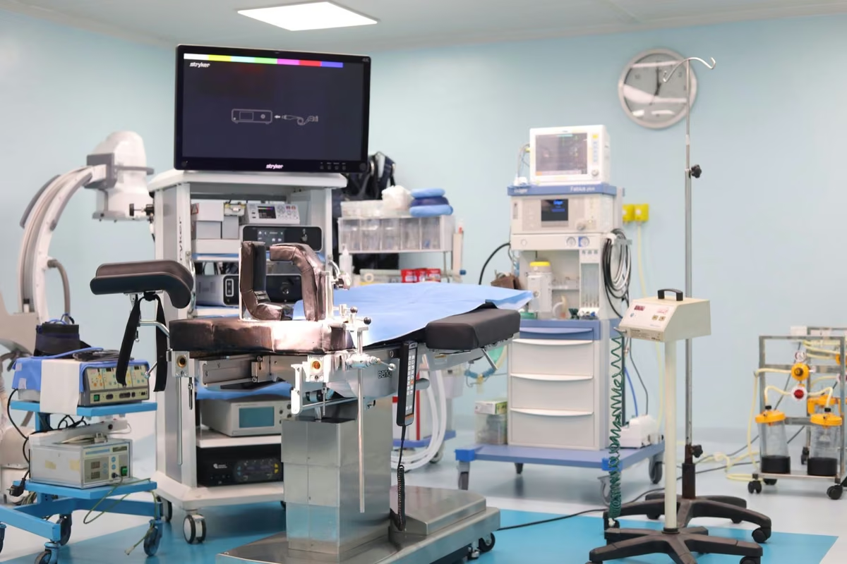
- This procedure is performed in the endoscopy unit under general anesthesia. If this has not already been done in the patient’s room, the nurse will insert an IV into a vein in the arm so that the anesthesiologist can administer the anesthesia through it.
- The anesthesiologist often inserts a tube into the airway to prevent breathing problems.
- During the exam, you will lie on your left side with a mouthpiece between your teeth and lips to prevent you from biting the endoscope.
- During the test, the oxygen level in your blood is measured continuously. This is done using a small device placed on your finger. You will also be given a pair of oxygen goggles to wear over your nose.
- In some cases, a dose of antibiotics is administered as a preventive measure during the procedure.
The Treatment
The endoscope is used to clearly delineate the lesion, which is then marked all around so it can be completely removed with a wide margin. Next, a thin needle is used to inject fluid between the muscle layer and the layer to be removed, allowing for safe manipulation. An incision is made around the lesion using a very fine electric scalpel. The lesion to be removed is then completely detached from the muscle layer using the fine blade. Finally, the wound is carefully inspected, and all blood vessels are cauterized with an electric current to prevent bleeding. The duration of the treatment varies depending on the size and location of the lesion: between 60 and 180 minutes. The removed lesion is preserved for subsequent microscopic examination.
After the examination
- After the procedure, you will be taken to the recovery room in the endoscopy unit. There, your blood oxygen levels will continue to be monitored.
- You may experience a scratchy throat for a while. You may also experience chest and/or stomach pain during the first few days.
- You may also experience bloating due to the carbon dioxide that was injected during the exam. Burping or passing gas can provide quick relief.
- After this procedure, you may be admitted to the hospital for a short time.
- On the day of the procedure, you may only drink water. Starting on the second day, you may generally begin eating liquid foods. After that, gradually transition to soft foods, and starting on the tenth day, you may eat normally.
- If you have a high fever, unbearable pain, difficulty swallowing, or bleeding (vomiting blood or rectal bleeding), you should contact the department or the doctor who treated you.
Risks
Bleeding
The procedure may cause a small blood vessel to be damaged. This is normal during the procedure and can almost always be stopped. Delayed bleeding between the first and 14th day is possible; this may manifest as vomiting blood or blood in the stool. If this occurs, a new endoscopy is needed to stop the bleeding. Delayed bleeding can occur in about 5 percent of procedures.
Perforation
This creates a small hole in the muscle layer, resulting in an open passage between the treated organ and the abdominal or thoracic cavity. This complication is rare and can occur in about 4 percent of procedures. In such cases, antibiotics are administered, and an attempt is usually made to close the hole.
Stenosis
During the healing process, this causes a narrowing of the treated organ, preventing food from passing through properly. This complication is very rare and can almost always be resolved by dilating the narrowing with a small balloon.